

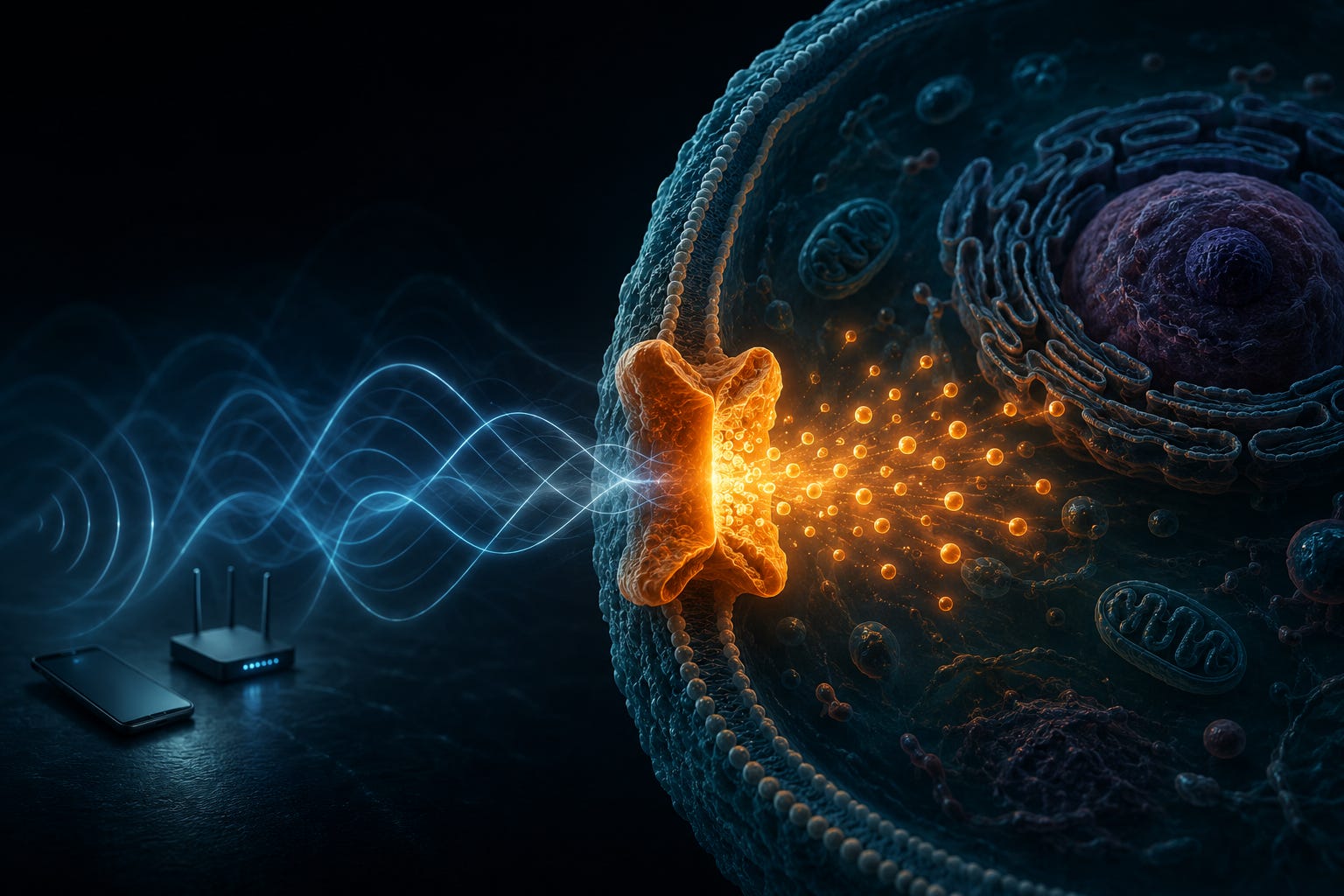
Part 1 covered what EMF is and why it matters. Part 2 showed you how to measure it. This piece answers the question both of those left hanging: how does a non-thermal electromagnetic field damage living tissue?
The answer involves a structure in your cell membranes so exquisitely sensitive to electrical forces that it amplifies them 7.2 million times. And the fact that this structure exists, that it’s been studied for decades, that we understand exactly how it works, is what makes the entire regulatory framework not just wrong but irrelevant. They’re measuring the wrong thing. Entirely. On purpose.
I should warn you - this article gets complicated. That’s because our bodies are incredibly complicated. Once you read this, you’ll understand just how naive we’re being by flooding our environment with electro-magnetic fields without the faintest understanding of how our bodies actually use electricity every second of every day to live. You might also get an inkling into why biological health and fertility are dropping so dramatically we’re looking at human extinction in a matter of just a few generations. This is serious stuff.
The Short Version (No Biology Degree Required)
If the science below makes your eyes glaze, here is the whole thing in plain English.
Your body runs on electricity. Not as a metaphor. Every heartbeat, every thought, every muscle twitch is triggered by tiny electrical signals shuttling charged particles in and out of your cells. To manage that, your cells are studded with microscopic gates that snap open and shut in response to the smallest change in electrical charge. They have to be exquisitely sensitive, because the signals they listen for are faint. Think of them as ears tuned to a whisper.
One of those gates controls calcium, the “go” button for a huge share of what your cells do. Because it matters so much, the gate is normally locked tight and only cracks open for a brief, controlled moment when the body says so.
Here is the problem. Precisely because these gates are built to detect the faintest electrical whisper, they are wildly oversensitive to the artificial electromagnetic fields pouring out of phones, routers, and cell towers. The physics works out to roughly a 7 million fold amplification: the force these fields put on the gate’s sensor is about seven million times stronger than what the rest of the cell feels. A whisper to you is a foghorn to the gate.
So the gate gets forced open when it shouldn’t. Calcium floods in. In small, brief doses that flood can actually heal, which is why medical devices use gentle pulses to help mend broken bones. But around the clock, day after day, the flood turns destructive. It drives inflammation, chemical damage, and, in animal studies, catastrophic brain cell death. Young rats exposed to cell tower levels of radiation lost roughly a third of their brain cells in four weeks. Give them a drug that plugs the calcium gate, and the damage almost entirely disappears. That is how we know the gate is the culprit.
Now for the complicated version:
The Gate
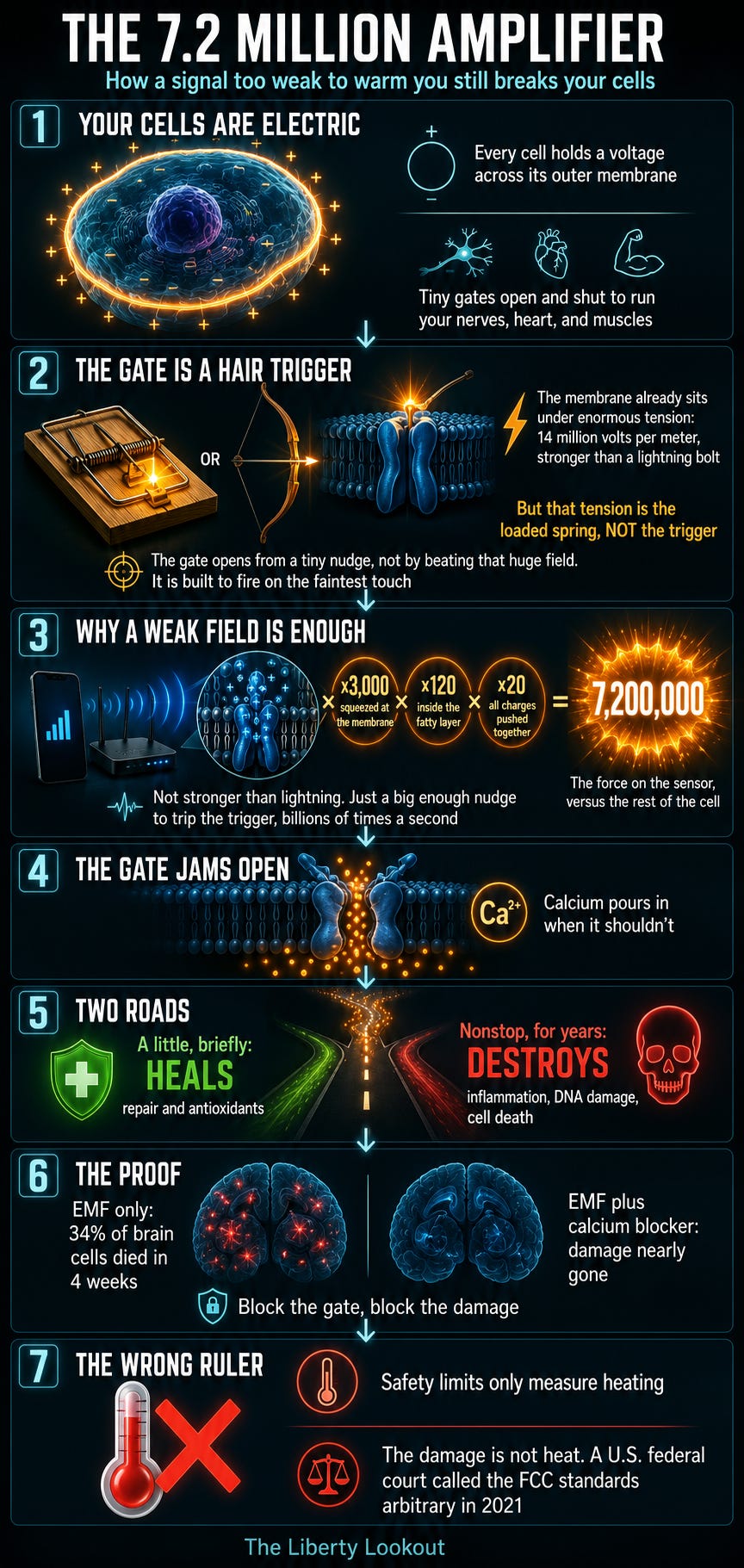
Every cell in your body maintains a careful balance of electrically charged particles (ions) between its interior and the fluid outside. The difference between these charges creates a voltage across the cell membrane, typically around -70 millivolts in a resting nerve cell. That’s not much. About one-twentieth of a single AA battery.
But “not much” is relative. The plasma membrane is only about 5 nanometers thick. That -70 millivolts spread across 5 nanometers works out to roughly 14 million volts per meter. The inside of a lightning bolt is about 3 million volts per meter. Your cells, right now, are maintaining electric field gradients that would make a thunderstorm jealous.
(The 14 million volts per meter is a static baseline, not the trigger. Think of the resting membrane as a heavy door held under enormous spring tension, balanced on a hair trigger. That huge tension is always there. It is not what opens the door. What opens the door is a tiny nudge to the trigger. In a real cell, the natural “nudge” is a small shift in membrane voltage, on the order of a few tens of millivolts, riding on top of that giant baseline. The sensor is tuned to detect that small change, not to be overpowered by the whole field. The system sits right at a threshold, poised to flip.)
Living in this intense field are proteins called voltage-gated calcium channels, or VGCCs. They sit embedded in the cell membrane like microscopic drawbridges, and their entire purpose is to detect changes in that membrane voltage and respond by opening or closing. When they open, calcium ions flood into the cell from outside, where the concentration of calcium is roughly 10,000 times higher than inside (Berridge et al., 1998).
Calcium is the most important signaling molecule in your body. It triggers neurotransmitter release. It initiates muscle contraction. It activates gene expression. It regulates cell division and cell death. Scientists call it the “universal second messenger” for good reason. Practically every critical cellular process uses calcium as its “go” signal.
(The “first messenger” is the signal that arrives from outside the cell: a hormone or neurotransmitter that docks onto a receptor on the cell surface but doesn’t actually enter the cell. Classic examples are adrenaline (epinephrine), glucagon, and neurotransmitters. The first messenger delivers the message to the cell, but it stays at the door. When it binds the surface receptor, that triggers the release of a second messenger inside the cell - calcium, cAMP, cGMP, IP3 - which relays the signal onward to the machinery that actually does the work. So calcium being a “second messenger” just means it’s the internal relay, not the original external signal.)
This is why cells keep intracellular calcium so absurdly low (around 100 nanomolar, or one ten-thousandth of the concentration outside). The gradient has to be steep because the signal has to be clear. A tiny controlled pulse of calcium entering the cell means “do this specific thing right now.” Uncontrolled flooding of calcium means catastrophe.
And the gatekeepers controlling that flood are VGCCs.
The Sensor
Here’s where the biology gets beautiful in a terrifying way.
Each VGCC is built around a large alpha-1 subunit protein that forms the actual channel pore. This protein contains four homologous domains (I through IV), each made of six membrane-spanning alpha helices labeled S1 through S6. The S5 and S6 helices from each domain arrange themselves to form the actual hole through which calcium flows.
But the business end, the part that matters for everything that follows, is the S4 helix in each domain. This is the voltage sensor.
The S4 helix carries positively charged amino acid residues (arginine and lysine) arranged in a repeating pattern. Each S4 helix typically carries 4 to 8 positive charges, and the VGCC has four of these sensor helices (one per domain). Adding up the functionally significant charges across all four voltage sensors gives you approximately 20 positive charges that must all be pushed in the same direction to open the gate.
Think of it like this: the VGCC is a lock that requires 20 tumblers to move simultaneously. When the voltage across the membrane shifts, the electrical force pushes all 20 charged groups in the S4 helices outward, the helices physically rotate and slide, and the channel opens. Calcium rushes in. The cell responds.
This is how your nervous system fires. This is how your heart beats. This is how muscles contract. Every thought you’re having while reading this sentence involves VGCCs opening and closing in precise, controlled patterns.
The system is extraordinarily sensitive by design. It has to be. Your entire electrochemical signaling apparatus depends on detecting tiny voltage shifts across the membrane and converting them into chemical signals. Evolution spent hundreds of millions of years optimizing this sensitivity.
Now consider what happens when you flood the environment with artificial electromagnetic fields.
The Amplifier
In 2013, Martin Pall, Professor Emeritus of Biochemistry and Basic Medical Sciences at Washington State University, published a paper in the Journal of Cellular and Molecular Medicine that laid out the evidence: EMFs act primarily through the activation of voltage-gated calcium channels.
He wasn’t the first to notice. Researchers had been documenting calcium increases in cells exposed to electromagnetic fields for decades. But Pall did something nobody else had done: he compiled all the studies in which calcium channel blocking drugs had been used to test whether VGCCs were the mechanism, and found that in study after study (two dozen of them), blocking calcium channels blocked the EMF effects. Different blockers targeting different types of calcium channels (L-type, T-type, N-type, P/Q-type) all produced protection. Each blocker is highly specific to calcium channels, making alternative explanations, as Pall put it, “highly unlikely.”
Then in 2015, he published a detailed biophysical analysis explaining why VGCCs are so extraordinarily sensitive to EMFs. The argument comes down to three multiplicative factors.
Factor 1: Membrane amplification (~3,000x). The plasma membrane has high electrical resistance. The fluids on either side of it (extracellular and intracellular) are good conductors. So when an external EMF passes through the body, the electric field moves charges easily through the conducting fluids but gets blocked at the membrane. This concentrates electrical forces perpendicular to the membrane surface. Even skeptics of non-thermal effects have acknowledged this produces roughly a 3,000-fold amplification of force on any structures sitting in the membrane.
Factor 2: Dielectric amplification (~120x). The voltage sensor’s charged groups sit inside the lipid bilayer of the membrane. Lipids have a dielectric constant similar to hydrocarbon solvents, which is low. The aqueous fluids surrounding the membrane have a dielectric constant roughly 120 times higher (water alone is ~40x higher than hydrocarbons, and the dissolved salts push it to roughly 2.5 to 3.5x higher than pure water). By Coulomb’s law, the force between electrical charges is inversely proportional to the dielectric constant of the medium between them. So the 20 charged groups on the voltage sensor, sitting in the low-dielectric lipid environment, experience electrical forces roughly 120 times greater than charges floating in the aqueous phase.
Factor 3: Coherent force on 20 charges (20x). An electromagnetic field produces a force in one direction at any given instant. All 20 positive charges on the voltage sensor get pushed the same way simultaneously. Random thermal energy, by contrast, is random in three dimensions. The probability of thermal energy spontaneously pushing all 20 charges in the same direction at the same instant is vanishingly small. EMFs do this every single cycle. At 2.4 GHz (WiFi frequency), that’s 2.4 billion times per second.
Multiply those three factors together:
3,000 × 120 × 20 = 7,200,000
The electrical force exerted on the VGCC voltage sensor by an external EMF is approximately 7.2 million times greater than the force that same field exerts on singly charged groups dissolved in the cytoplasm.
In other words - the structure that evolution designed to be the most electrically sensitive component in your cell is experiencing forces from your WiFi router that are 7.2 million times stronger than what the rest of the cell feels.
This is why Pall concluded that EMF fields “6 to 7 orders of magnitude” below the threshold needed to produce heating can still activate VGCCs and produce biological effects. The thermal threshold is irrelevant. It’s like trying to determine the biological effects of sound by measuring whether it heats the eardrum. Your eardrum doesn’t work by getting hot. It works by vibrating. VGCCs don’t respond to heat. They respond to voltage.
The Two Roads
So EMFs activate VGCCs. Calcium floods in. What happens next?
This is where the biology reveals something both elegant and disturbing. The calcium doesn’t just do one thing. It triggers two competing pathways, and which one dominates depends on how much calcium enters and for how long.
The therapeutic pathway runs like this: a modest, brief increase in intracellular calcium activates a protein called calmodulin. Calmodulin, with calcium bound to it, then activates the constitutive nitric oxide synthases (eNOS and nNOS), which produce nitric oxide (NO). The NO activates soluble guanylyl cyclase, producing cGMP, which activates protein kinase G. This cascade also stimulates a transcription factor called Nrf2, which cranks up the cell’s antioxidant defenses: superoxide dismutases, glutathione synthesis enzymes, peroxynitrite scavengers. The whole system is protective and regenerative.
This is why pulsed electromagnetic field (PEMF) therapy actually works for bone healing and tissue repair. Controlled, brief, low-intensity pulses produce modest calcium increases that kick the therapeutic pathway into gear. Orthopedic surgeons have been prescribing PEMF devices for non-union fractures since the 1970s.
In 2012, researcher Arthur Pilla published a striking study showing that pulsed EMFs produced a nearly 3-fold increase in calcium/calmodulin-dependent nitric oxide synthesis, and the response was nearly instantaneous, occurring in less than 5 seconds. The speed of the response was itself revealing: the calcium increase had to occur almost immediately after EMF exposure, consistent with direct activation of VGCCs (which open and close in microseconds), not some slow thermal process.
The pathological pathway runs differently. When calcium entry is excessive and sustained (chronic exposure, high intensity, or both), the system overloads. Too much NO is produced. Simultaneously, the mitochondria, stressed by excessive calcium, produce superoxide. Nitric oxide and superoxide react to form peroxynitrite (ONOO⁻), one of the most destructive molecules in biology.
Peroxynitrite attacks DNA. It oxidizes lipids. It damages proteins. It depletes tetrahydrobiopterin (BH4), a cofactor that nitric oxide synthase needs to function properly. And here’s where it gets vicious: when BH4 is depleted, the NOS enzymes themselves become “uncoupled” and start producing more superoxide instead of nitric oxide, feeding the very cycle that’s destroying the cell. Peroxynitrite also activates NF-κB, the master inflammatory transcription factor, driving chronic production of inflammatory cytokines.
The final twist: these two pathways inhibit each other. Nrf2 (the protective pathway) raises enzymes that scavenge peroxynitrite. Peroxynitrite (the destructive pathway) oxidizes the components the therapeutic pathway needs to function. Once the peroxynitrite pathway gains the upper hand, it progressively dismantles the cell’s ability to recover.
Brief, controlled calcium increases can heal you. Chronic, excessive calcium increases will destroy you. And the regulatory agencies measuring safety by checking whether your tissue gets warm are capturing exactly none of this.
The Proof: Block the Channel, Block the Damage
Science has a simple test for whether a proposed mechanism is real: block the proposed mechanism and see if the effect disappears.
Pall’s 2013 review compiled two dozen studies in which researchers blocked calcium channels with specific drugs and then exposed cells or animals to EMFs. The result was consistent across all of them: blocking the calcium channel blocked the EMF effect.
The most dramatic example comes from El-Swefy and colleagues at Zagazig University in Egypt. In a 2008 study published in the Journal of Applied Biomedicine, they exposed young rats to cell phone base station radiation (81 milligauss) for just 2 hours per day.
After four weeks, the results were devastating. The EMF-exposed rats showed progressive oxidative damage, prolonged elevated calcium, pro-inflammatory markers (TNFα and CRP), and widespread brain cell death.
34% of the brain cells died in four weeks from an exposure level comparable to living near a cell tower.
But a second group of rats received amlodipine, a calcium channel blocker, alongside the same EMF exposure. In this group, all 11 measured brain changes and all 4 observed behavioral changes were substantially reduced. Amlodipine “restored the EMF-induced apoptosis in brain to near normal.”
Consider what this means. If EMFs damaged tissue through heating, blocking calcium channels wouldn’t make any difference. You don’t prevent a burn by blocking calcium channels. The fact that a calcium channel blocker almost completely prevents EMF-induced brain damage is definitive evidence that the damage pathway runs through calcium channels.
And it wasn’t just amlodipine. Across the studies Pall reviewed, five different classes of calcium channel blockers (targeting different types and different binding sites on VGCCs) all blocked various EMF effects. L-type blockers acting at two different sites. T-type blockers. N-type blockers. P/Q-type blockers. All effective. All highly specific to calcium channels. The probability that all of these specific drugs happen to block EMF damage through some mechanism other than calcium channel blockade is, in Pall’s words, “highly unlikely.”
18 Roads to Alzheimer’s
In 2022, Pall published what may be the most alarming paper of his career: “Low Intensity Electromagnetic Fields Act via Voltage-Gated Calcium Channel (VGCC) Activation to Cause Very Early Onset Alzheimer’s Disease: 18 Distinct Types of Evidence” in Current Alzheimer Research.
The calcium hypothesis of Alzheimer’s disease isn’t new or controversial. For almost 25 years, researchers have built a robust case that excessive intracellular calcium produces each of the major features of Alzheimer’s: amyloid-beta plaques, hyperphosphorylated tau protein, neurofibrillary tangles, synaptic dysfunction, neuroinflammation, and neuronal death through both apoptosis and destructive autophagy. The Alzheimer’s Association itself published a framework in 2017 integrating calcium dysregulation into a comprehensive theory of Alzheimer’s pathogenesis.
Pall’s contribution was connecting this directly to electromagnetic fields. His 18 types of evidence include:
The mechanistic evidence: EMFs activate VGCCs. VGCC activation floods cells with calcium. Excessive calcium drives both the calcium signaling pathway and the peroxynitrite/oxidative stress/inflammation pathway. Both pathways produce Alzheimer’s-specific pathology. A vicious cycle develops where amyloid-beta protein and intracellular calcium amplify each other.
The genetic evidence: Human genetic studies show that people with elevated VGCC activity (genetic variants that make their calcium channels more active) have increased Alzheimer’s incidence.
The pharmacological evidence: Calcium channel blockers reduce Alzheimer’s risk in clinical populations.
The epidemiological evidence: Twelve occupational exposure assessments found that people with higher occupational EMF exposures have higher Alzheimer’s incidence. A 2019 meta-analysis across multiple studies found a weighted relative risk of 1.33 (33% higher Alzheimer’s incidence) for occupational EMF exposure. Some studies suggest EMFs shorten the normal 25-year latency period of the disease.
The temporal evidence: The age of onset of Alzheimer’s has been dropping over the past two decades, corresponding precisely to the massive increase in wireless communication EMF exposures. Recent studies now report Alzheimer’s cases in people aged 30 to 40.
The animal evidence: This is where it gets truly disturbing.
El-Swefy’s 2008 study (the one with amlodipine) showed universal massive neurodegeneration in young rats from base station radiation. 34% brain cell death in four weeks. Just 2 hours per day.
Jiang et al. (2013) exposed 2-month-old rats to electromagnetic pulses and found apparent universal Alzheimer’s effects by 20 months of age. In human terms, that’s roughly equivalent to 42-year-olds developing universal Alzheimer’s.
A follow-up experiment with daily EMF pulse exposure found Alzheimer’s markers developing at 10 months, as described in Pall’s 2022 analysis. In human terms: 21-year-olds with Alzheimer’s.
“Any of these may produce the ultimate nightmare,” Pall wrote, “extremely early onset Alzheimer’s Disease.”
Beyond Calcium: The Whole System
VGCCs get the most attention because the calcium they control triggers the most dramatic downstream effects. But here’s the uncomfortable broader truth: VGCCs aren’t the only voltage-gated ion channels in your body.
Voltage-gated sodium channels, potassium channels, and chloride channels all share the same fundamental architecture. They all have S4 voltage-sensing helices packed with positively charged residues. They all sit in the plasma membrane, subject to the same amplification factors (membrane concentration of forces, dielectric constant differences, coherent multi-charge activation).
As early as 2002, Dimitris Panagopoulos and colleagues at the University of Athens published a biophysical model predicting that EMFs would cause forced oscillation of free ions near the voltage sensors of all voltage-gated ion channels, not just calcium. Their model predicted that the ion vibrations would irregularly gate these channels, disrupting normal function. Follow-up work in 2021 confirmed this prediction and linked the mechanism to oxidative stress and DNA damage.
The research has focused primarily on VGCCs because calcium produces the most obvious biological effects and because calcium channel blockers are widely available and well-characterized drugs. But the biophysics of the voltage sensor applies to the entire family of voltage-gated ion channels (VGICs). This means EMFs aren’t just disrupting calcium signaling. They’re potentially disrupting the entire electrochemical signaling system of the body: nerve impulse propagation (sodium channels), cardiac rhythm (sodium, potassium, and calcium channels), muscle function (all of the above), and cellular homeostasis (potassium and chloride channels).
The body is, at its most fundamental level, an electrical machine. Every nerve signal, every heartbeat, every muscle twitch, every thought runs on precisely controlled ion flows through voltage-gated channels. We’ve built a civilization that saturates these systems with artificial electromagnetic fields billions of times stronger than the natural background they evolved in.
And nobody is measuring the relevant forces.
The Wrong Ruler
The entire global regulatory framework for electromagnetic field safety is built on a single metric: Specific Absorption Rate (SAR). SAR measures the rate at which the body absorbs electromagnetic energy as heat, in watts per kilogram.
This made a kind of intuitive sense in the 1960s, when the main concern was military radar operators standing too close to high-powered transmitters and literally cooking their tissues. Heat is a real danger at sufficiently high power levels. Nobody disputes this.
But SAR is a thermodynamic measure. It tells you about energy absorbed as heat. It tells you nothing about the electrical forces acting on voltage-gated ion channels. And those forces, as we’ve now seen, are amplified 7.2 million times at the voltage sensor relative to the thermal effects on the bulk of the cell.
It’s as if we tested car safety exclusively by measuring whether the engine catches fire, concluded that all cars driving under 200 mph are safe because no engines ignited, and ignored the entire concept of crash protection. “The engine didn’t catch fire at 120 mph, so 120 mph is safe.” Nobody asks about the humans inside, because the testing framework wasn’t designed for them.
In 2021, the U.S. Court of Appeals for the DC Circuit told the FCC exactly this. The court found that the FCC’s decision to maintain its existing safety standards was “arbitrary and capricious” because the agency had failed to address evidence of non-thermal biological effects. The court specifically noted the FCC’s failure to respond to evidence about harm to children, the environment, and long-term exposures at non-thermal levels.
The FCC’s response since that ruling? More or less nothing. The standards haven’t changed.
Here’s the core of the problem: the question was never “do non-thermal EMFs heat tissue?” That answer has always been “no, not at typical exposure levels,” and it has always been the wrong question. The question is: “do non-thermal EMFs exert biologically relevant forces on structures designed to detect electrical changes?”
The answer, supported by decades of research, two dozen calcium channel blocker studies, animal models showing universal neurodegeneration, epidemiological data showing elevated Alzheimer’s risk, and a biophysical analysis showing a 7.2-million-fold amplification factor, is yes.
What This Means for You
The entire “safe until it heats you” framework collapses once you understand VGCCs. Non-thermal fields don’t need to heat tissue to activate calcium channels, because the voltage sensor amplifies incoming electromagnetic forces by millions. Pulsed fields (which is what every wireless communication device produces) are more biologically active than continuous fields, a fact that was recognized in safety standards as far back as the 1960s and then conveniently forgotten as the wireless industry grew.
The mechanism also explains why “smarter” devices may be worse. Each generation of wireless technology uses more complex pulsation patterns to carry more data. More complex pulsation means more voltage transitions per second. More voltage transitions means more VGCC activations. 5G uses beam-forming, massive MIMO, and extreme pulse modulation. The data throughput goes up. So does the biological damage.
The practical steps in Part 2 (measuring, removing sources, hardwiring) aren’t precautionary theater. They’re directly responsive to a documented mechanism. Every wireless device you remove from your sleeping area is one less source of chronic VGCC activation. Every smart device you replace with a wired alternative is one less stream of pulsed electromagnetic forces acting on the most sensitive electrical structures in your body.
Part 5 will follow the calcium downstream: into the immune system, where chronic VGCC activation doesn’t just damage neurons. It degrades the terrain.
Sources
- Pall, M.L. (2013). “Electromagnetic fields act via activation of voltage-gated calcium channels to produce beneficial or adverse effects.” Journal of Cellular and Molecular Medicine, 17(8), 958-965. PMC3780531
- Pall, M.L. (2015). “Scientific evidence contradicts findings and assumptions of Canadian Safety Panel 6.” Reviews on Environmental Health, 30(2), 103-116. De Gruyter
- Pall, M.L. (2022). “Low Intensity Electromagnetic Fields Act via Voltage-Gated Calcium Channel (VGCC) Activation to Cause Very Early Onset Alzheimer’s Disease: 18 Distinct Types of Evidence.” Current Alzheimer Research, 19(2), 119-132. PubMed 35114921
- Pilla, A.A. (2012). “Electromagnetic fields instantaneously modulate nitric oxide signaling in challenged biological systems.” Biochemical and Biophysical Research Communications, 426(3), 330-333. PubMed 22940137
- El-Swefy, S. et al. (2008). “Calcium channel blockade alleviates brain injury induced by long term exposure to an electromagnetic field.” Journal of Applied Biomedicine, 6(3), 153-163. DOI: 10.32725/jab.2008.019
- Jiang, D.P. et al. (2013). “Electromagnetic pulse exposure induces overexpression of beta amyloid protein in rats.” Archives of Medical Research, 44(3), 178-184. PubMed 23523687
- Catterall, W.A. (2010). “Ion Channel Voltage Sensors: Structure, Function, and Pathophysiology.” Neuron, 67(6), 915-928. PMC2950829
- Panagopoulos, D.J. et al. (2002). “Mechanism for action of electromagnetic fields on cells.” Biochemical and Biophysical Research Communications, 298(1), 95-102. PubMed 12379225
- Panagopoulos, D.J. et al. (2021). “Human-made electromagnetic fields: Ion forced-oscillation and voltage-gated ion channel dysfunction, oxidative stress and DNA damage.” International Journal of Oncology, 59(5), 92. PubMed 34617575
- Gunnarsson, L.G. & Bodin, L. (2019). “Occupational Exposures and Neurodegenerative Diseases—A Systematic Literature Review and Meta-Analyses.” Int. J. Environ. Res. Public Health, 16(3), 337. MDPI
- Berridge, M.J. et al. (1998). “Calcium — a life and death signal.” Nature, 395, 645-648. PubMed 9790183
- Alzheimer’s Association Calcium Hypothesis Workgroup (2017). “Calcium Hypothesis of Alzheimer’s disease and brain aging.” Alzheimer’s & Dementia, 13(2), 178-182. PubMed 28061328
- EurekAlert! (2022). “EMFs, calcium and Alzheimer’s disease: A closer link.” Press release